- Citas Centro Médico de Caracas: Lunes, Miercoles y Viernes. Pulse el botón Agende una Cita
- Sistema de citas en linea exclusivo para Centro Medico de Caracas en San Bernardino
- Citas CMDLT: Jueves. llamar al 0212-9496243 y 9496245
- Las Emergencias son atendidas en CMDLT previa coordinacion personal al 04142708338
- Proveedor Seguros Mercantil y Sudeban

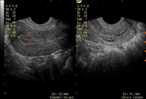
Intracavitary Transvaginal Sonography (TVUS) allow us to obtain the first pregnancy images of an unequivocal intrauterine pregnancy when there is one of sufficient gestational age to be seen: this is the most modern and more sensitive method for early pregnancy evaluation of early pregnancy compared to the traditional abdominal scan.
Embryo ultrasound
Week 3: Before the 4 weeks of gestation, the presence of intrauterine pregnancy can not be detected by Echo, this is because the product of conception is less than 1 mm and that is the range of resolution of modern equipment via transvaginal In this period, modern pregnancy tests are positive even though we do not see anything by Eco. Embryologically there is already an embryo of similar size to this point “.” It has the shape of an oval disk divided into three layers that are forming the first sketches of organs, especially the Central Nervous System. In this period is when defects such as Spina Bifida or Anencephaly are formed that we can only begin to see 6 to 7 weeks later; in fact, many of the problems that we will detect later are being developed at this moment.
This stage refers to the so-called ” biochemical pregnancy ” because despite there being a positive pregnancy test in no other way, neither by physical examination nor even by ultrasound, an objective fact can be put in evidence that there is actually a pregnancy in progress.
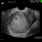
Week 4: First evidence of pregnancy manifested by the presence of a small 2 mm sac. of diameter (Gestational sac) inside which it is practically impossible to detect any element. Although nothing is seen ultrasound, there is an embryo growing rapidly inside the sac. By this time the spine is in the process of formation. If there is a spina bifida problem, its origin is occurring at this moment.
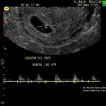
Week 5 : As the days go by, the Gestational Sack grows and inside it begins to see a small ‘cupped ball’ (Vitelina Vesicle) followed by the appearance, next to it, of a white “granite” of 1 to 2 mm . (the embryo) The embryo continues to grow and towards the end of the 5th week a small and rapid heartbeat begins to be felt inside: we see the first manifestation of life of the human embryo, the Cardiac Activity (the heart, which only has 0.5 to 1 mm already it can be heard using Doppler technology). The normal frequency must be greater than 80 beats to consider a good prognosis.
Week 6: The structures of the 5 week are easier to evaluate, the cardiac activity is much easier to see and measure. You begin to notice a bulge in one of the ends that will be the head. It measures approximately 4.5 mm.
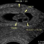
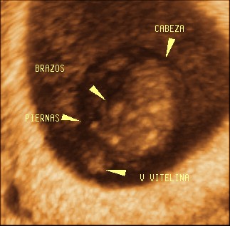
Week 7: A little larger, 9.2 mm. The head is remarkable and you can see some brain structures, the heart is bigger and you begin to see four points that will be the extremities. The umbilical cord begins to be visualized. The Vitelina Vesicle is easily appreciated.
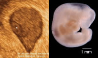
Week 8: Now it measures about 15 mm. The head has grown and new brain structures have appeared, the heart is bigger and the 4 limbs are more evident. DOES IT MOVE!!! with small embryonic movements (but movements you will only feel them from week 16 to 20). The cord is easily seen and you can see how it reaches the future placenta. 3D Eco.
Week 9 : Our embryo measures 22 mm. The division of head and body begins to be noticed (the development of the neck began), all visible structures are larger and more defined, the four limbs have been lengthened a bit and formed “palettes” but no fingers are seen yet. His movements are more frequent and notorious. 3D Eco.
In the following video you can see the different stages of embryo-fetal development according to Carnegie’s stadiums. Material copyright: The Multi-Dimensional Human Embryo. Bradley R. Smith .
References
The Fetal Medicine Foundation Series, The 11-14 week scan. 2007
Embryology. Langman Ed. Panamericana 5th Ed.
The Visible Embryo, http://www.visembryo.com
Human Reproduction Update 1996, Vol. 3, No. 1 pp. 3-23 European Society for Human Reproduction and Embryology. Morphological and molecular characteristics of human fetuses between Carnegie stages 7 and 23: developmental stages in the post-implantation embryo . LMHarkness1 and DTBair.
Transvaginal transducer
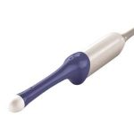
This is the transducer used until week 13, for intravaginal use, to obtain better resolution and quality of images in early pregnancy. This is a 5-9 MHz General Electric Transvaginal model with 3D / 4D capability
