- Citas Centro Médico de Caracas: Lunes, Miercoles y Viernes. Pulse el botón Agende una Cita
- Sistema de citas en linea exclusivo para Centro Medico de Caracas en San Bernardino
- Citas CMDLT: Jueves. llamar al 0212-9496243 y 9496245
- Las Emergencias son atendidas en CMDLT previa coordinacion personal al 04142708338
- Proveedor Seguros Mercantil y Sudeban

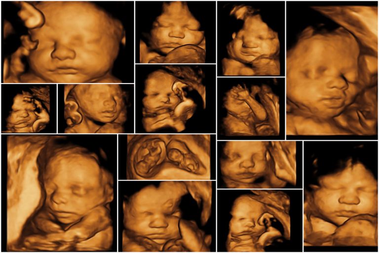
Three-dimensionality has been included in healthcare medicine since the mid-1990s, 3D Ecosonography began in the world around the year 2000 and has become so popular that a large part of pregnant women has undergone a study of this type. The advantage of this method lies in the easy comprehension of the images obtained by people not trained in imaging and the possibility of clearly documenting the findings made in each study. In addition, 3D attractiveness has made many patients approach a high-level diagnostic study since most of the ultrasound practitioners who practice 3D’s are highly trained in prenatal diagnosis
In our experience we have noticed that the best results are obtained between weeks 24 and 29. The study is easily done, the anatomy is clearly seen, the circulatory Doppler study begins to have relevance and we obtain 3D images from medium to high quality in more 90% of the cases. The study begins by assessing the vitality, position and number of fetuses; then, we make the measurements to verify growth, we continue with the advanced anatomical description, the Doppler study and finally we make the 3D shots of fetal parts, especially the face and genitals. This is a study that has become routine even in the absence of formal indications and we have noticed that a favorable result translates into a pleasant sense of family tranquility.
The origin of the 3D image is based on 3 or more cuts obtained by the system, orthogonal cuts or multiplanar image, these are cuts obtained by the evaluated dimensions (2) and other cuts calculated or integrated by the equipment. This allows us to obtain images of complex organs such as the brain and the heart that we would not otherwise be able to obtain. This resource is what really makes 3D an unparalleled tool.
Ultrasound 5D
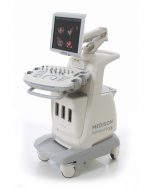
Currently there is 5D ultrasound ( Samsung healthcare ), much more sensitive and with more multiplanar cuts, better resolution, speed in real time and new diagnostic resources. 4D images of the face and fetal parts are much more detailed
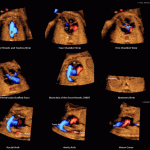
This is an example of 5D ultrasound, not only get up to 9 different cuts but that exposes them in motion and with color Doppler capability
In Venezuela there are not many 5D due to the cost, however, with the available 4D technology our diagnostic sensitivity is comparable.
Click on this image to see an example of real 4D video
Cover Image: Hi-Care General Hospital Ltd. Dhaka
3D realities
1.- The color of the photos is artificial.
2.- The study is done as a normal ultrasound in gray colors
3.- The difference of the study is the capacity of the operator and the technical resources of the latest generation equipment
4.- The best time to conduct a study of Second Level 3D/4D Doppler is between 24 and 29 weeks .
5.- We added the 4D study to the week 18-20, when we can, because the moving images are simply excellent
6.- 3D images can not always be obtained: maternal obesity, location of the placenta and variants of the fetal position can make this an impossible event (10-15% of cases)
7.- It is not convenient to try to obtain 3D images at all costs because, being a part of the study that has no diagnostic value, the maternal discomforts or the risk of overstimulating the gravid uterus are not justified. I have as a rule not to exceed 30-35 minutes, but this varies according to the criteria of the operator.
8.- 5D ultrasound provides incredible advantages for the diagnosis of complex anomalies in organs such as the fetal heart

