- Citas Centro Médico de Caracas: Lunes, Miercoles y Viernes. Pulse el botón Agende una Cita
- Sistema de citas en linea exclusivo para Centro Medico de Caracas en San Bernardino
- Citas CMDLT: Jueves. llamar al 0212-9496243 y 9496245
- Las Emergencias son atendidas en CMDLT previa coordinacion personal al 04142708338
- Proveedor Seguros Mercantil y Sudeban

The Obstetric Ultrasound equipment uses the same Radar technology: the emission of sound waves at an inaudible frequency (in MHz). These waves, when mechanically rebounding in the tissues, generate moving images of the visible structures of the baby, the classic gray-colored images that are the basis of the study and the way in which the advanced diagnoses are made, there is no irradiation. The added color is artificial either in the Doppler or in the 3D/4D image of the baby.
Glossary and definitions
Sonography, Echosonography, Ultrasonography: all synonyms. It involves the use of equipment emitting and receiving high frequency “sounds” (ultrasonic, not audible by the human ear, (more than 20KHz, usually between 3.5 and 9.5 MHz) that collect the “echoes” of the structures studied and translates them in visible images, so we assemble complex, even three-dimensional, images of the object under study, in our case, the baby.In Latin countries we use the term “Eco, Ultrasound” a lot to refer to Ultrasound studies (US). to use any of these terms interchangeably.
Transducers : these are the pieces of the Ultrasound Equipment that the doctor holds in their hands and which they place on the patient’s body to obtain the image of the desired organ or system. There are Surface, such as the one that is placed on your abdomen (US Transabdominal or USTA) and Endocavitarios that serve to make studies through natural cavities such as the vagina (US Transvaginal or USTV). In early pregnancies (under 13 weeks) the transvaginal route allows us to obtain a high resolution image of a baby (embryo or fetus) from 3 to 85 mm in length that otherwise would be impossible to evaluate
Obstetric Ecosonography : is the exclusive study of the pregnant uterus and its content. Special emphasis is placed on the evaluation of the Fetus (Anatomy and Fetal Physiology) and its Annexes (Placenta, Umbilical Cord and Amniotic fluid).
Doppler echography : this is a specialized study that aims to assess the circulatory status of the fetus and its mother. Through its use we can detect early the potential risk of fetal growth disorders, compromise of fetal well-being and hypertensive diseases in the mother. It requires sophisticated and expensive equipment. Through Doppler we can hear the amplified sound of the heartbeats and see through artificial colors the direction of the Fetal and Maternal blood flow.
Second level Obstetric Ecosonography (Level II) : it is also known as Advanced Ultrasound because it involves the use of high resolution equipment managed by specialists with extensive experience in the detection of structural and functional disorders of the fetus. This is a study aimed at ruling out fetal anomalies or determining the origin of an anomaly previously detected. In experienced hands it could detect up to 95% of fetal problems depending on the affected organ or system. Otherwise, a normal study without visible anomalies greatly guarantees that your baby is healthy.
Genetic Ecosonography : specialized study with high resolution equipment that aims to rule out the presence of markers (small structural organic changes) that are associated with chromosomal problems, genetic syndromes and complex congenital anomalies. There are two types: First Trimester (11 to 13.6 weeks) and Second Trimester (18 to 23 weeks).
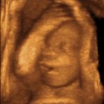
Three-dimensional Ecosonography (US-3D) : it is the obtaining of three-dimensional images of the whole baby (before the 14th week of pregnancy) or parts of it (face, genitals, limbs, etc). It only involves obtaining 3D images without reference to the rest of the study. Many people confuse the US-3D with the study of Level II when they are in fact different studies: the truth is that in most of the serious centers where the US-3D is practiced the study carried out incorporates the study of Level II, Doppler evaluation and obtaining 3D images; this is why some confusion has been generated in this regard. Doppler 3-4D devices are the most advanced and expensive in the market.
Tetradimensional Ecosonography (US-4D) : with the latest generation of commercial equipment it is possible to obtain three- dimensional images of a baby in real time movement (4D); thus, the fourth dimension refers to the moving image of a baby in 3 dimensions. You can watch a short 4D ultrasound video here .
Ecosonography 5D (US-5D): is a new technology from Samsung that generates volumes, 3D images and multiple automated cuts that greatly facilitate the diagnosis of static and dynamic fetal organs. The cardiac evaluation allows simultaneous cuts with everything necessary to rule out or diagnose congenital heart disease; likewise, it facilitates the evaluation of the CNS (Central Nervous System), skeletal and fetal genetic evaluation
Reach
Is such an advanced diagnosis adequate?
The “danger” of these very diligent studies is that they could detect with a fetal problem: it sounds illogical but there are people who can not tolerate the anguish of knowing beforehand that there is a problem, but most patients prefer to know it to make decisions and prepare . Fortunately most of the babies are healthy and the parents are grateful for a study that ruled out a lot of diseases
How often is there a structural finding?
We found some positive marker or anatomical variation in only 1 to 2 of every 100 patients studied (1-2%). Most of these findings are considered normal anatomical variants, one case goes to other studies including Amniocentesis and a truly pathological case is rarely found.
Functional disorders?
These are found with a much higher frequency. Approximately 5 to 10 cases per 100 patients will present some functional disorder (delayed fetal growth, fetal distress, circulatory alterations, decreased Amniotic fluid, etc.) detected by Doppler and other ultrasound tests. Most of them will be mild and occasionally we will find some severe case in which the US will be the decisive weapon to determine the best moment for the termination of pregnancy.
Limitations of the method
Ultrasound studies are not perfect methods and may fail to detect maternal or fetal problems.
The technological advances and the skills and knowledge of the operator are determinant in the quality of the study, if there is any detectable problem the operator will detect it, but it will depend on these 4 factors:
- Equipment: the most modern and hi-tech ones are designed to obtain images of great quality and detail due to its high resolution and imaging resources but their cost limit the availability, and studies are usually more expensive for the patient.
- Operator: nothing would be achieved with the best equipment available if your operator is not qualified to carry out a quality study. The preparation and diagnostic capacity of the specialist is the element that most often determines the quality of a study
- Patient: in patients with large adipose panniculus (central obesity) the team loses a lot of diagnostic capacity.
- Fetus: there are conditions that do not manifest or are so small that they are not detectable. Decreased Amniotic Liquid content severely limits the quality of the study
Ob sonogaphy is not game

We add to our daily obstetric practice tools that would make our predecessors pale with envy: we have equipment that allows us to sniff inside the baby’s “house” and evaluate its development, growth and well-being; we can see it on the outside and inside, know with absolute certainty the blue or pink color (metaphor about fetal sex) of his future clothes or if we will have to buy everything double; in short, we see the baby in its natural environment, previously secret, to discover little by little its wonderfully small but complex universe.
Although fun and sometimes informal, the study by ultrasound is a serious medical act that requires a lot of preparation and responsibility in its execution. The result of the study must be reflected in a detailed report endorsed by a certified specialist in the area.
Although a single case of fetal damage attributable to the medical use of ultrasound has never been reported, in more than 30 years of use, we take great care not to exert much or prolonged pressure or excessive uterine manipulation. That is to say, once the necessary information has been obtained, the study is suspended because there is no point in continuing to bother the baby; This is especially true in study 3-4D: if the baby does not show his face in a prudential time, the study is declared completed, there will be time to see it when it is born!